
WHAT IS PTERYGIUM?
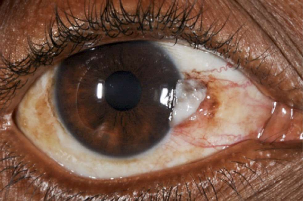
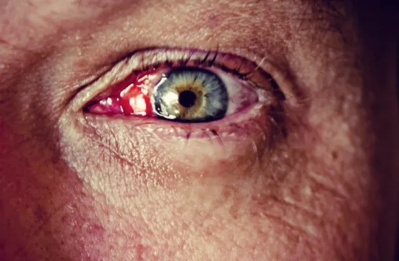
Pterygium is a raised, pinkish, wing-shaped fleshy growth on the conjunctiva of the eye extending towards the cornea. Ninety percent of the cases occur on the nasal side.
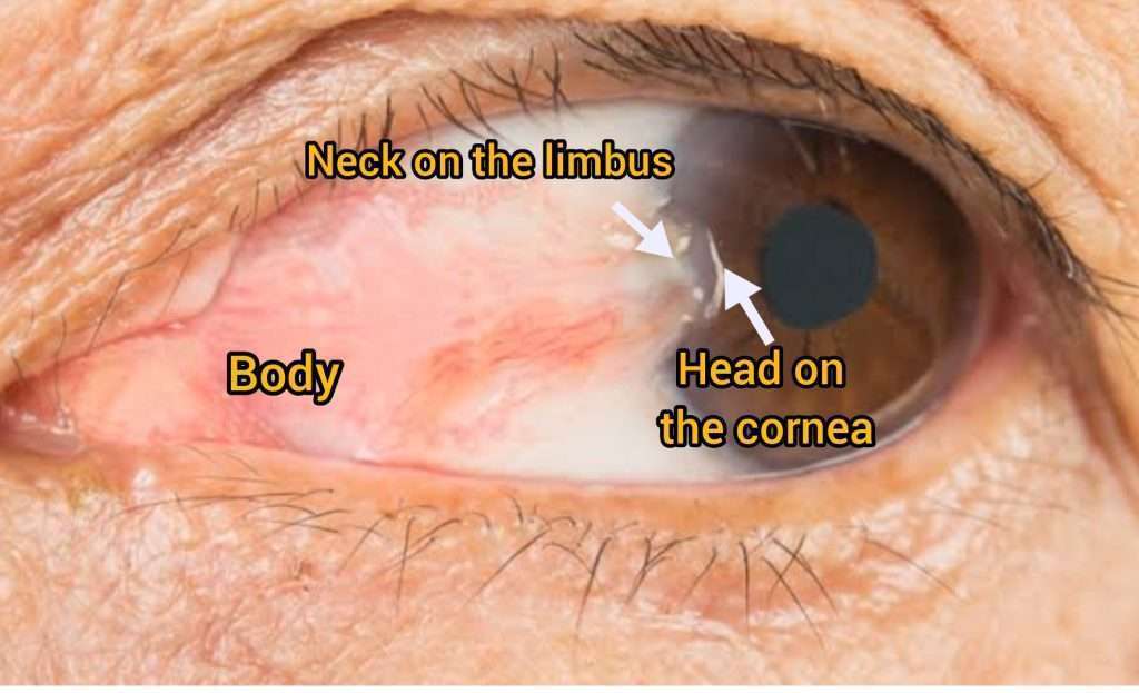
Essentially, it consists of three identifiable parts: an apex (head), a neck, and a body. With the head extending towards the cornea and the base towards the inner part of the eye.
WHY IS PTERYGIUM MOSTLY NASAL?
1. The transmission of UV rays by internal reflection in the cornea may lead to a concentration of UV irradiation at the nasal limbus, hence the high incidence of nasal pterygium.
2. Nasal pterygium may be the result of the normal flow of tears from outwards to inwards carrying with them any dust or sand particles or fine foreign bodies, thereby triggering an inflammatory response.
WHAT IS SURFER’S EYE?
Experiencing direct sunlight and reflection from the water for a prolonged period of time puts surfers at risk for pterygium. Hence Pterygium is often known as Surfer’s eye.
CAUSES:
Pterygium has no definitive cause, but following are the contributing risk factors:
- UV rays: UV rays can cause inflammation in the conjunctiva, oxidative stress, and even loss of limbal stem cells, resulting in pterygiums.
- People living near the equator of the earth due to more sunlight exposure
- People living in hot, dry environment
- People spending more time outdoors such as farmers, surfers, fishermen etc.
- Genetic
SYMPTOMS:
In most cases, pterygium is asymptomatic, but it can sometimes lead to discomfort that needs medical attention.

- Redness
- Burning
- Irritation
- Watering
- Unclear vision if encroaching the cornea
- Foreign body sensation
IS PTERYGIUM CANCEROUS?
Pterygium is a noncancerous or benign fibrovascular growth on the white part of the eye growing towards the cornea.
Sometimes an eye cancer called OSSN can look like pterygium. The OSSN has 3 clinical forms, papillary (raised), gelatinous (gel like) and leukoplakia (white lesion)
Therefore examination by an eye doctor is very important for proper diagnosis.
WHAT IS NOT A PTERYGIUM?
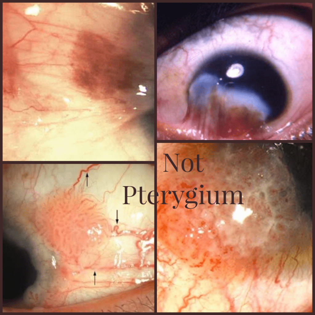
- The most common location for Pterygium is in the nasal area, with only 5-10% occurring temporally .Pterygium in other regions like superiorly, or inferiorly is highly unlikely and can actually be symblepharon due to injury or inflammation.
- Excessive pigmentation
- Very irregular surface
- Large tortuous vessels on the lesion
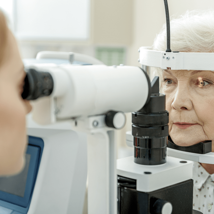
DIAGNOSIS:
The diagnosis of pterygium is clinically made using a slit lamp examination in the doctor’s office. It is not necessary to perform special tests to diagnose pterygium.
EYEDROPS FOR PTERYGIUM REMOVAL
There are no eyedrops to treat pterygium. Surgery is the only treatment option. Pterygium is usually asymptomatic but sometimes can cause redness, irritation, watering and foreign body sensation in which case a doctor can prescribe some anti-inflammatory eye drops such as steroids. It is important to use these drops only under the doctor’s supervision.
WHAT ARE THE INDICATIONS FOR SURGERY?
Surgery is usually not needed when pterygiums are asymptomatic and do not cause much discomfort, however it can be surgically removed in following cases:
1. Threatening the vision by one of the following mechanisms:
- Covering the visual axis
- Threatening the visual axis
- Corneal curvature problems causing astigmatism: If the pterygium is over 3mm it may cause some astigmatism and if it is over 3.5mm (extending beyond the middle of the pupil in a typical cornea) it may cause > 1d astigmatism.
2. Eye movement restriction
3. Documented progression by doctor or by patient.
4. Symptoms of irritation, watering, recurrent redness
WHAT IS PTERYGIUM SURGERY?
Common technique used today is simple excision of the pterygium and covering the exposed area with a garft using a glue or sutures. The grafts are either conjunctival autograft, limbal autograft or amniotic membrane graft.
In order to prevent recurrences, antifibrotic drops are used before or after the surgery. Some surgeons also employ beta irradiation on the surface as a preventative measure.
Graft is then placed on the area where the pterygium was excised, either with glue or sutures.
The excimer laser may be used to smoothen out irregularities on the cornea when the pterygium is very large and involves the cornea.
After pterygium surgery the patient may have redness and irritation for about 1-2 weeks. During the recovery phase it is important to use the drops as prescribed by the doctor.
WHAT ARE THE COMPLICATIONS OF SURGERY
Like any other surgery a pterygium excision surgery also has some complications
1. Recurrence: Main problem with surgery is recurrence. It is common for recurrences to occur within 12 months of surgery. Recurrent pterygium is different from the primary pterygium. In severe cases it may adhere to the surrounding tissue making their excision extremely challenging.
2. Subconjunctival hemorrhage
3. Swelling in the graft
4. Displacement or loss of graft
5. Retraction
6. Granuloma, and
7. Less commonly corneal melt, perforation.
WHAT TO DO IF YOU HAVE PTERYGIUM AND CATARACT
It is usually recommended to have pterygium surgery first if it is causing vision problems. It takes three months for the eye curvature to stabilize after pterygium excision, and then measurements are taken to calculate the IOL for cataract surgery.
PREVENTION IS BETTER THAN CURE
Protecting your eyes from ultraviolet rays, dust, wind and allergies is very important to prevent pterygium. Consider implementing the following:

- Protect your eyes from the sun by wearing UV-protective sunglasses
- Wide-brimmed hats provide additional protection for your eyes
- Use lubricating eye drops to wash off any inflammatory mediators from dust, wind and sun that can cause pterygium