Is 20/20 vision the ideal vision? What exactly does having healthy eyes mean?
Regular eye exams are necessary for everyone as they can help avoid future vision issues. It’s critical to select the best eye test for your needs out of the many available options. Your doctor or optometrist can provide you with the best advice regarding which test you need.
Here are 11 tests available to assess the eye health.
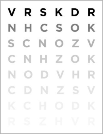
1. VISUAL ACUITY TEST
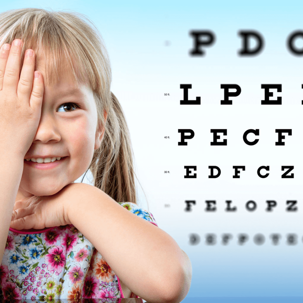
This test evaluates both distance and near vision. A normal vision is described as 20/20 or 6/6. Simply put, it means that you can make out an object that is 20 feet or 6 metres away from you. If you have 20/30 or 6/9 vision, you can see objects from 20 feet or 6 metres away that a person with normal vision can see from 30 feet or 9 metres away. This indicates that you have less vision than normal.
Near vision is measured with a small handheld chart held at 35cm by the patient. The size of text in this chart decreases from the top to the bottom.
While having 20/20 or 6/6 vision indicates that a person has good central vision, it does not imply that their visual function is perfect or ideal.
In addition to visual acuity, there are other aspects of overall eye health to take into account. These factors include peripheral vision, stereopsis or depth perception, colour vision, contrast sensitivity, and others.
2. REFRACTION TEST
Refraction determines your prescription for eyeglasses.
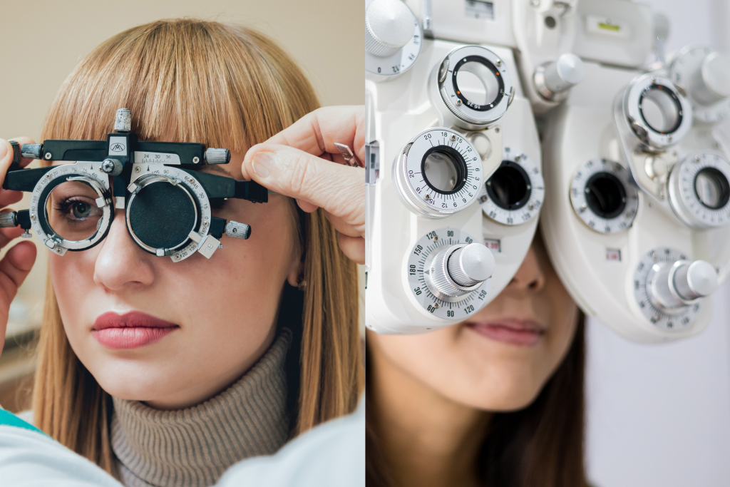
You either wear a trial frame which has a slot for keeping the lenses in front of your eyes, or you sit in a chair with a special device (called a phoropter or refractor) attached to it. Phoropter comes with lenses of different strengths that can be moved into the field of view.
You look through the instrument or lenses, focusing on an eye chart kept at a distance of 20 ft (6-meter). In this test, each eye is tested separately.
The optometrist uses the information you provide to determine whether your eyes are healthy, you don’t require a prescription for vision correction, or you are nearsighted, farsighted, or have astigmatism.
3. CONTRAST SENSITIVITY:
Contrast sensitivity refers to a person’s capacity to recognise low-contrast objects in their environment, such as a street sign on a dark street or a cloudy day, or the height of a sidewalk step at night.
Using low-contrast images, contrast sensitivity testing detects subtle vision changes that would otherwise remain undetected by visual acuity testing.
Numerous eye conditions, including diabetes, cataracts, glaucoma, lazy eye, optic nerve conditions, etc., have an effect on patients’ contrast sensitivity.
4. COLOUR VISION:
Red, green, and blue are the three primary colours that the human eye can distinguish. Trichromatic vision, also known as trichromacy, is the standard form of colour vision for humans.
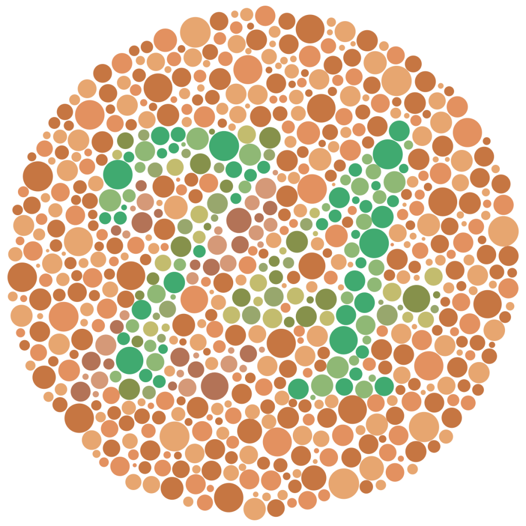
Ishihara colour deficiency tests can diagnose red-green deficiency, the most common colour deficiency.
The Ishihara test is a book which consists of a collection of plates with a range of coloured and sized dots on them. A person with normal colour vision can easily read the numbers contained within these dots. A person with a red-green deficiency experiences difficulty in reading the plates correctly.
5. BINOCULARITY:
Synoptophore is a device for testing binocular vision. Binocular vision has three grades:
Grade 1 is simultaneous perception. Both eyes can simultaneously perceive two images, one formed on each retina.
Grade 2 is fusion. Both eyes can combine their visual information to produce a single vision; and
Grade 3, stereopsis, also known as depth perception or 3D viewing.
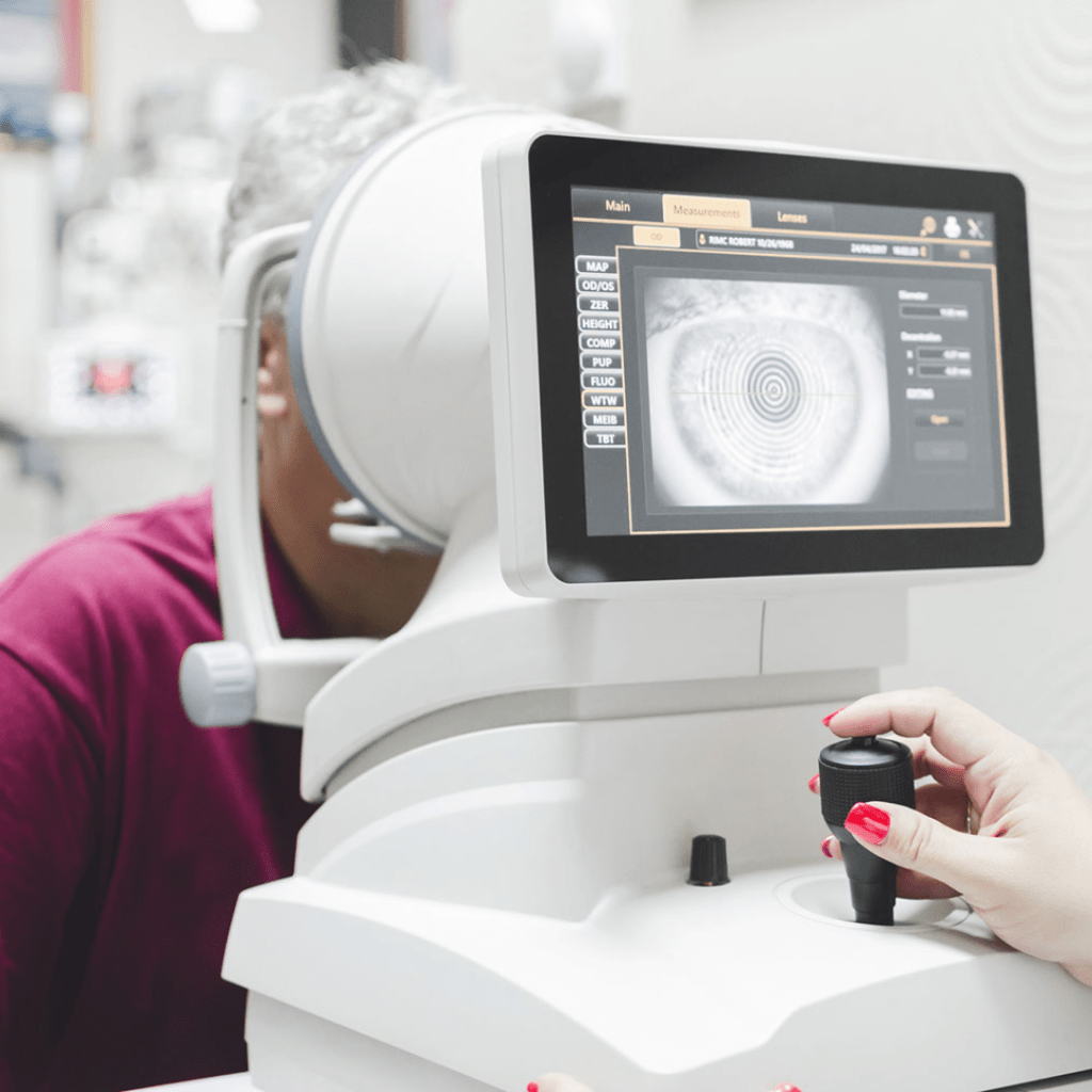
6. KERATOMETRY TEST
This test measures the cornea’s curvature, the eye’s outer layer. The shape of your cornea affects how you perceive and reflect light.
Astigmatism is a condition that occurs when a person’s cornea has steep or elongated curves. Optometrists use keratometry tests to detect astigmatism and other issues with the cornea’s curvature, such as keratoconus.
7. PERIPHERAL VISUAL FIELD TEST
Even though we tend to focus our attention on whatever we are looking at directly, our peripheral vision allows us to see things at the edges of our field of vision.
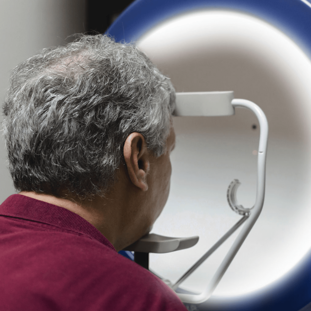
Visual field tests measure how far we can see. Automated perimetry is the most popular kind of peripheral visual field test.
You look into a special machine and focus on a spot in the centre. You must press the button each time you notice a light flash in your peripheral vision while maintaining a constant focus on the centre.
An optometrist can use this test to measure the size of your visual field and identify any blind spots in your peripheral vision.
8. INTRAOCULAR PRESSURE MEASUREMENT
An intraocular pressure (IOP) test measures the force or pressure produced by the fluid in your eyes.
An abnormally high level of eye pressure is a sign of glaucoma.
The procedure involves having you sit before a non-contact intraocular measurement device that pokes your open eye with air for a brief period of time.
Based on how your eye responds to the air puff, the machine then measures your eye pressure.
There is another type of IOP measurement that is more accurate. The eyes are numbed with anesthetic eye drops then stained with fluorescein dye. The applanation tonometer, a specialised tool used in this test, gently touches your eye to measure the internal pressure.
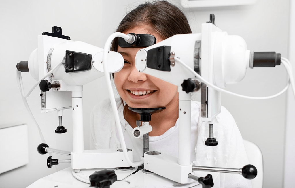
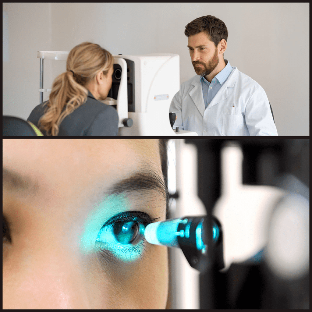
9. SLIT LAMP EXAMINATION:
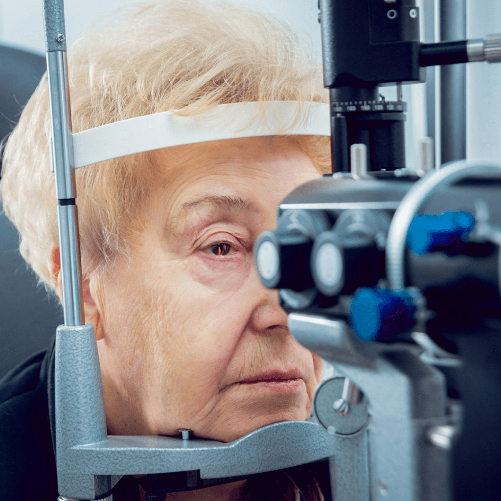
Slit lamp is microscopy allows your ophthalmologist to get a better look at the various structures inside the eye and on the front of the eye. It is a crucial tool for assessing your eye health and identifying eye diseases.
This examination can also be used to examine the optic nerve. Glaucoma is an eye condition that slowly kills the optic nerve and impairs vision if not identified and treated early.
10. DRY EYE TEST:
The quantity and quality of the tear film that covers our eyes play a critical role in preserving the ocular surface’s health.

The routine dry eye tests conducted in clinics measure these aspects of your tear production.
1.Amount or output: Schirmer Test
2. Quality: TBUT or tear break-up time
3. Osmolarity
11. DILATED FUNDUS EXAMINATION
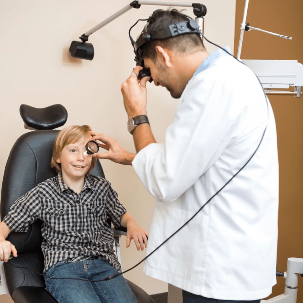
While slit lamps can see inside the eye, indirect ophthalmoscopes give a wider view.
Your ophthalmologist will use dilating eye drops to dilate (widen) your pupils to achieve this. The retina and optic nerve inside the eye are carefully examined for any kind of abnormality.
This test can detect retinal pathologies like retinal tears, inherited retinal diseases like retinitis pigmentosa, and age-related macular degeneration.
QUICK SUMMARY:
A 20/20 vision is not always indicative of an ideal vision. That, along with good contrast sensitivity, color vision, binocularity, the field of vision, and the health of both internal and external structures, all contribute to good vision. If you haven’t had a routine eye exam yet, make an appointment with your eye doctor now.